
Journal of Structural Biology
FEI’s direct electron detector developments: Embarking on a revolution in cryo-TEM
Publication date: Available online 9 October 2015
Source:Journal of Structural Biology
Author(s): Maarten Kuijper, Gerald van Hoften, Bart Janssen, Rudolf Geurink, Sacha De Carlo, Matthijn Vos, Gijs van Duinen, Bart van Haeringen, Marc Storms
In early 2011 FEI Company launched the “Falcon”, its first commercial direct electron detector product intended for application in 3-D electron microscopy in the life sciences. In this paper we discuss the principle of direct electron detection and its implementation in Falcon cameras. We describe the signal formation in the sensor and its impact on the detection quantum efficiency (DQE) of the sensor. Insights into the signal formation led us to improved camera designs. Three significant improvements are discussed. (1) Back thinning of the sensor. This is implemented in the second-generation Falcon (Falcon 2), where the sensor thickness is reduced to 50μm, and in the latest generation Falcon 3 detector with further back-thinning down to 30μm. (2) The introduction of electron counting, a signal processing technology implemented in Falcon 3. (3) Dose fractionation mode, which allows the user to access intermediate results during the illumination of the sample.
Categories: Journal Articles
Simultaneous determination of sample thickness, tilt, and electron mean free path using tomographic tilt images based on Beer–Lambert law
Publication date: Available online 9 October 2015
Source:Journal of Structural Biology
Author(s): Rui Yan, Thomas J. Edwards, Logan M. Pankratz, Richard J. Kuhn, Jason K. Lanman, Jun Liu, Wen Jiang
Cryo-electron tomography (cryo-ET) is an emerging technique that can elucidate the architecture of macromolecular complexes and cellular ultrastructure in a near-native state. Some important sample parameters, such as thickness and tilt, are needed for 3-D reconstruction. However, these parameters can currently only be determined using trial 3-D reconstructions. Accurate electron mean free path plays a significant role in modeling image formation process essential for simulation of electron microscopy images and model-based iterative 3-D reconstruction methods; however, their values are voltage and sample dependent and have only been experimentally measured for a limited number of sample conditions. Here, we report a computational method, tomoThickness, based on the Beer–Lambert law, to simultaneously determine the sample thickness, tilt and electron inelastic mean free path by solving an overdetermined nonlinear least square optimization problem utilizing the strong constraints of tilt relationships. The method has been extensively tested with both stained and cryo datasets. The fitted electron mean free paths are consistent with reported experimental measurements. The accurate thickness estimation eliminates the need for a generous assignment of Z-dimension size of the tomogram. Interestingly, we have also found that nearly all samples are a few degrees tilted relative to the electron beam. Compensation of the intrinsic sample tilt can result in horizontal structure and reduced Z-dimension of tomograms. Our fast, pre-reconstruction method can thus provide important sample parameters that can help improve performance of tomographic reconstruction of a wide range of samples.
Categories: Journal Articles
A fast cross-validation method for alignment of electron tomography images based on Beer–Lambert law
Publication date: Available online 9 October 2015
Source:Journal of Structural Biology
Author(s): Rui Yan, Thomas J. Edwards, Logan M. Pankratz, Richard J. Kuhn, Jason K. Lanman, Jun Liu, Wen Jiang
In electron tomography, accurate alignment of tilt series is an essential step in attaining high-resolution 3D reconstructions. Nevertheless, quantitative assessment of alignment quality has remained a challenging issue, even though many alignment methods have been reported. Here, we report a fast and accurate method, tomoAlignEval, based on the Beer–Lambert law, for the evaluation of alignment quality. Our method is able to globally estimate the alignment accuracy by measuring the goodness of log-linear relationship of the beam intensity attenuations at different tilt angles. Extensive tests with experimental data demonstrated its robust performance with stained and cryo samples. Our method is not only significantly faster but also more sensitive than measurements of tomogram resolution using Fourier shell correlation method (FSCe/o). From these tests, we also conclude that while current alignment methods are sufficiently accurate for stained samples, inaccurate alignments remain a major limitation for high resolution cryo-electron tomography.
Categories: Journal Articles
Automated batch fiducial-less tilt-series alignment in Appion using Protomo
Publication date: Available online 9 October 2015
Source:Journal of Structural Biology
Author(s): Alex J. Noble, Scott M. Stagg
The field of electron tomography has benefited greatly from manual and semi-automated approaches to marker-based tilt-series alignment that have allowed for the structural determination of multitudes of in situ cellular structures as well as macromolecular structures of individual protein complexes. The emergence of complementary metal-oxide semiconductor detectors capable of detecting individual electrons has enabled the collection of low dose, high contrast images, opening the door for reliable correlation-based tilt-series alignment. Here we present a set of automated, correlation-based tilt-series alignment, contrast transfer function (CTF) correction, and reconstruction workflows for use in conjunction with the Appion/Leginon package that are primarily targeted at automating structure determination with cryogenic electron microscopy.
Categories: Journal Articles
Mineral-bearing vesicle transport in sea urchin embryos
Publication date: Available online 9 October 2015
Source:Journal of Structural Biology
Author(s): Netta Vidavsky, Admir Masic, Andreas Schertel, Steve Weiner, Lia Addadi
Sea urchin embryos sequester calcium from the sea water. This calcium is deposited in a concentrated form in granule bearing vesicles both in the epithelium and in mesenchymal cells. Here we use in vivo calcein labeling and confocal Raman spectroscopy, as well as cryo-FIB-SEM 3D structural reconstructions, to investigate the processes occurring in the internal cavity of the embryo, the blastocoel. We demonstrate that calcein stained granules are also present in the filopodial network within the blastocoel. Simultaneous fluorescence imaging and Raman spectroscopy show that these granules do contain a calcium mineral. By tracking the movements of these granules, we show that the granules in the epithelium and primary mesenchymal cells barely move, but those in the filopodial network move long distances. We could however not detect any unidirectional movement of the filopodial granules. We also show the presence of mineral containing multivesicular vesicles that also move in the filopodial network. We conclude that the filopodial network is an integral part of the mineral transport process, and possibly also for sequestering calcium and other ions. Although much of the sequestered calcium is deposited in the mineralized skeleton, a significant amount is used for other purposes, and this may be temporarily stored in these membrane-delineated intracellular deposits.
Categories: Journal Articles
Reassessment of MxiH subunit orientation and fold within native Shigella T3SS needles using surface labelling and solid-state NMR
Publication date: Available online 6 October 2015
Source:Journal of Structural Biology
Author(s): Joeri Verasdonck, Da-Kang Shen, Alexander Treadgold, Christopher Arthur, Anja Böckmann, Beat H. Meier, Ariel J. Blocker
T3SSs are essential virulence determinants of many Gram-negative bacteria, used to inject bacterial effectors of virulence into eukaryotic host cells. Their major extracellular portion, a ∼50nm hollow, needle-like structure, is essential to host cell sensing and the conduit for effector secretion. It is formed of a small, conserved subunit arranged as a helical polymer. The structure of the subunit has been studied by electron cryomicroscopy within native polymers and by solid-state NMR in recombinant polymers, yielding two incompatible atomic models. To resolve this controversy, we re-examined the native polymer used for electron cryomicroscopy via surface labelling and solid-state NMR. Our data show the orientation and overall fold of the subunit within this polymer is as established by solid-state NMR for recombinant polymers.
Categories: Journal Articles
Analysis of distinct molecular assembly complexes of keratin K8 and K18 by hydrogen–deuterium exchange
Publication date: Available online 3 October 2015
Source:Journal of Structural Biology
Author(s): Aiswarya Premchandar, Anna Kupniewska, Krzysztof Tarnowski, Norbert Mücke, Monika Mauermann, Magdalena Kaus-Drobek, Aleksander Edelman, Harald Herrmann, Michał Dadlez
Keratins are intermediate filament (IF) proteins that form complex filament systems in epithelial cells, thus serving as scaffolding elements and mechanical stress absorbers. The building blocks of keratin IFs are parallel coiled-coil dimers of two distinct sequence-related proteins distinguished as type I and type II keratins. To gain more insight into their structural dynamics, we resorted to hydrogen–deuterium exchange mass spectrometry of keratins K8 and K18, which are characteristic for simple epithelial cells. Using this powerful technique not employed with IFs before, we mapped patterns of protected versus unprotected regions in keratin complexes at various assembly levels. In particular, we localized protein segments exhibiting different hydrogen exchange patterns in tetramers versus filaments. We observed a general pattern of precisely positioned regions of stability intertwining with flexible regions, mostly represented by the non-α-helical segments. Notably, some regions within the coiled-coil domains are significantly more dynamic than others, while the IF-consensus motifs at the end domains of the central α-helical “rod” segment, which mediate the “head-to-tail” dimer–dimer interaction in the filament elongation process, become distinctly more protected upon formation of filaments. Moreover, to gain more insight into the dynamics of the individual keratins, we investigated the properties of homomeric preparations of K8 and K18. The physiological importance of keratins without a partner is encountered in both pathological and experimental situations when one of the two species is present in robust excess or completely absent, such as in gene-targeted mice.
Categories: Journal Articles
Structural and biochemical insights into the DNA-binding mode of MjSpt4p:Spt5 complex at the exit tunnel of RNAPII
Publication date: Available online 2 October 2015
Source:Journal of Structural Biology
Author(s): Gongrui Guo, Yongxiang Gao, Zhongliang Zhu, Debiao Zhao, Zhihong Liu, Huihao Zhou, Liwen Niu, Maikun Teng
Spt5 (NusG in bacteria) is the only RNA polymerase-associated factor known to be conserved in all three domains of life. In archaea and eukaryotes, Spt5 associates with Spt4, an elongation factor that is absent in bacteria, to form a functional heterodimeric complex. Previous studies suggest that the Spt4:Spt5 complex interacts directly with DNA at the double-stranded DNA exit tunnel of RNA polymerase to regulate gene transcription. In this study, the DNA-binding ability of Spt4:Spt5 from the archaeon Methanocaldococcus jannaschii was confirmed via nuclear magnetic resonance chemical shift perturbation and fluorescence polarization assays. Crystallographic analysis of the full-length MjSpt4:Spt5 revealed two distinct conformations of the C-terminal KOW domain of Spt5. A similar alkaline region was found on the Spt4:Spt5 surface in both crystal forms, and identified as double-stranded DNA binding patch through mutagenesis-fluorescence polarization assays. Based on these structural and biochemical data, the Spt4:Spt5-DNA binding model was built for the first time.
Categories: Journal Articles
The giant keyhole limpet radular teeth: a naturally-grown harvest machine
Publication date: Available online 2 October 2015
Source:Journal of Structural Biology
Author(s): Tina Ukmar-Godec, Gregor Kapun, Paul Zaslansky, Damien Faivre
The limpet radula is a feeding organ, which contains more than 100 rows of teeth. During their growth the teeth mature and advance in position along the radula. The simpler doccoglossan radulae operate by grinding rocky substrates, extracting the algae by rasping and scraping with the teeth functioning as shovels. Less is known about the rhipidoglossan radulae, used as rakes or brooms that brush and collect loose marine debris. This type of radula is found in the giant keyhole limpet (Megathura crenulata). The large size of this organism suggests that the rhipidoglossan radula entails a technological superiority for Megathura crenulata in its habitat. The structure and function of the radulae teeth have however not been reported in detail. Using a combination of 2D and 3D microscopy techniques coupled with amino acid analysis and X-ray scattering, we reveal the working components of Megathura crenulata’s radula. It is characterized by numerous marginal teeth surrounding a pair of major hook-like lateral teeth, two pairs of minor lateral teeth and a large central tooth. The mature major lateral teeth show pronounced signs of wear, which gradually increase towards the very front end of the radula and are evidence for scraping. An abrupt change in the amino acid composition in the major lateral teeth and the concurrent formation of a chitinous fiber-network mark the onset of tooth maturation. In comparison to the simpler rock-scraping doccoglossate limpets, the radula of Megathura crenulata forms an elaborate feeding apparatus, which can be seen as a natural harvest machine.
Categories: Journal Articles
Molecular events during the early stages of aggregation of GNNQQNY: An all atom MD simulation study of randomly dispersed peptides
Publication date: Available online 2 October 2015
Source:Journal of Structural Biology
Author(s): Alka Srivastava, Petety V. Balaji
This study probes the early events during lag phase of aggregation of GNNQQNY using all atom MD simulations in explicit solvent. Simulations were performed by varying system size, temperature and starting configuration. Peptides dispersed randomly in the simulation box come together early on in the simulation and form aggregates. These aggregates are dynamic implying the absence of stabilizing interactions. This facilitates the exploration of alternate arrangements. The constituent peptides sample a variety of conformations, frequently re-orient and re-arrange with respect to each other and dissociate from/re-associate with the aggregate. The size and lifetime of aggregates vary depending upon the number of inter-peptide backbone H-bonds. Most of the aggregates formed are amorphous but crystalline aggregates of smaller size (mainly 2-mers) do appear and sustain for varying durations of time. The peptides in crystalline 2-mers are mostly anti-parallel. The largest crystalline aggregate that appears is a 4-mer in a single sheet and a 4-, 5-, or 6-mer in double layered arrangement. Crystalline aggregates grow either by the sequential addition of peptides, or by the head-on or lateral collision-adhesion of 2-mers. The formation of various smaller aggregates suggests the polymorphic nature of oligomers and heterogeneity in the lag phase.
Categories: Journal Articles
Cover 2 - Editorial Board
Publication date: October 2015
Source:Journal of Structural Biology, Volume 192, Issue 1
Source:Journal of Structural Biology, Volume 192, Issue 1
Categories: Journal Articles
Table of Contents / barcode
Publication date: October 2015
Source:Journal of Structural Biology, Volume 192, Issue 1
Source:Journal of Structural Biology, Volume 192, Issue 1
Categories: Journal Articles
The REC domain mediated dimerization is critical for FleQ from Pseudomonas aeruginosa to function as a c-di-GMP receptor and flagella gene regulator
Publication date: October 2015
Source:Journal of Structural Biology, Volume 192, Issue 1
Author(s): Tiantian Su, Shiheng Liu, Kang Wang, Kaikai Chi, Deyu Zhu, Tiandi Wei, Yan Huang, Liming Guo, Wei Hu, Sujuan Xu, Zong Lin, Lichuan Gu
FleQ is an AAA+ ATPase enhancer-binding protein that regulates both flagella and biofilm formation in the opportunistic pathogen Pseudomonas aeruginosa. FleQ belongs to the NtrC subfamily of response regulators, but lacks the corresponding aspartic acid for phosphorylation in the REC domain (FleQR, also named FleQ domain). Here, we show that the atypical REC domain of FleQ is essential for the function of FleQ. Crystal structure of FleQR at 2.3Å reveals that the structure of FleQR is significantly different from the REC domain of NtrC1 which regulates gene expression in a phosphorylation dependent manner. FleQR forms a novel active dimer (transverse dimer), and mediates the dimerization of full-length FleQ in an unusual manner. Point mutations that affect the dimerization of FleQ lead to loss of function of the protein. Moreover, a c-di-GMP binding site deviating from the previous reported one is identified through structure analysis and point mutations.
Categories: Journal Articles
Investigating interactions of the Bacillus subtilis spore coat proteins CotY and CotZ using single molecule force spectroscopy
Publication date: October 2015
Source:Journal of Structural Biology, Volume 192, Issue 1
Author(s): Huiqing Liu, Daniela Krajcikova, Zhe Zhang, Hongda Wang, Imrich Barak, Jilin Tang
Spores formed by Bacillus subtilis are surrounded by a protective and multilayered shell, termed the coat, which grants the spores resistance to various environmental stresses and facilitates spore germination. The spore coat consists of more than seventy different proteins, arranged into at least four distinct structural layers: the undercoat, inner coat, outer coat and crust. However, how these proteins, especially the morphogenetic proteins, interact to establish the organized, functional coat layers remains poorly understood. CotY and CotZ as the components of the crust, play a morphogenetic role in the crust assembly around the spore. In this study, the single molecule force spectroscopy was used to investigate the interaction and dynamics between CotY and CotZ at the single-molecule level. The results show that homotypic interactions of CotY and CotZ and the heterotypic interaction between CotY and CotZ exist. Furthermore, the dissociation kinetics of the complexes were studied by monitoring the relationship between the unbinding forces and the loading rates at different pulling velocities. In this way, a series of kinetic parameters regarding the three different complexes were obtained. It revealed the strong interactions between CotY and CotZ, CotY and CotY, and a relatively weak interaction of CotZ and CotZ.
Categories: Journal Articles
Structure of neurotropic adeno-associated virus AAVrh.8
Publication date: October 2015
Source:Journal of Structural Biology, Volume 192, Issue 1
Author(s): Sujata Halder, Kim Van Vliet, J. Kennon Smith, Thao Thi Phuong Duong, Robert McKenna, James M. Wilson, Mavis Agbandje-McKenna
Adeno-associated virus rhesus isolate 8 (AAVrh.8) is a leading vector for the treatment of neurological diseases due to its efficient transduction of neuronal cells and reduced peripheral tissue tropism. Toward identification of the capsid determinants for these properties, the structure of AAVrh.8 was determined by X-ray crystallography to 3.5Å resolution and compared to those of other AAV isolates. The capsid viral protein (VP) structure consists of an αA helix and an eight-stranded anti-parallel β-barrel core conserved in parvoviruses, and large insertion loop regions between the β-strands form the capsid surface topology. The AAVrh.8 capsid exhibits the surface topology conserved in all AAVs: depressions at the icosahedral twofold axis and surrounding the cylindrical channel at the fivefold axis, and three protrusions around the threefold axis. A structural comparison to serotypes AAV2, AAV8, and AAV9, to which AAVrh.8 shares ∼84%, ∼91%, and ∼87% VP sequence identity, respectively, revealed differences in the surface loops known to affect receptor binding, transduction efficiency, and antigenicity. Consistent with this observation, biochemical assays showed that AAVrh.8 is unable to bind heparin and does not cross-react with conformational monoclonal antibodies and human donor serum directed against the other AAVs compared. This structure of AAVrh.8 thus identified capsid surface differences which can serve as template regions for rational design of vectors with enhanced transduction for specific tissues and escape pre-existing antibody recognition. These features are essential for the creation of an AAV vector toolkit that is amenable to personalized disease treatment.
Categories: Journal Articles
X-ray structural and molecular dynamical studies of the globular domains of cow, deer, elk and Syrian hamster prion proteins
Publication date: October 2015
Source:Journal of Structural Biology, Volume 192, Issue 1
Author(s): Pravas Kumar Baral, Mridula Swayampakula, Adriano Aguzzi, Michael N.G. James
Misfolded prion proteins are the cause of neurodegenerative diseases that affect many mammalian species, including humans. Transmission of the prion diseases poses a considerable public-health risk as a specific prion disease such as bovine spongiform encephalopathy can be transferred to humans and other mammalian species upon contaminant exposure. The underlying mechanism of prion propagation and the species barriers that control cross species transmission has been investigated quite extensively. So far a number of prion strains have been characterized and those have been intimately linked to species-specific infectivity and other pathophysiological manifestations. These strains are encoded by a protein-only agent, and have a high degree of sequence identity across mammalian species. The molecular events that lead to strain differentiation remain elusive. In order to contribute to the understanding of strain differentiation, we have determined the crystal structures of the globular, folded domains of four prion proteins (cow, deer, elk and Syrian hamster) bound to the POM1 antibody fragment Fab. Although the overall structural folds of the mammalian prion proteins remains extremely similar, there are several local structural variations observed in the misfolding-initiator motifs. In additional molecular dynamics simulation studies on these several prion proteins reveal differences in the local fluctuations and imply that these differences have possible roles in the unfolding of the globular domains. These local variations in the structured domains perpetuate diverse patterns of prion misfolding and possibly facilitate the strain selection and adaptation.
Categories: Journal Articles
Structural characterization and modeling of the Borrelia burgdorferi hybrid histidine kinase Hk1 periplasmic sensor: A system for sensing small molecules associated with tick feeding
Publication date: October 2015
Source:Journal of Structural Biology, Volume 192, Issue 1
Author(s): William J. Bauer, Amit Luthra, Guangyu Zhu, Justin D. Radolf, Michael G. Malkowski, Melissa J. Caimano
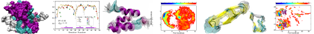
Two-component signal transduction systems are the primary mechanisms by which bacteria perceive and respond to changes in their environment. The Hk1/Rrp1 two-component system (TCS) in Borrelia burgdorferi consists of a hybrid histidine kinase and a response regulator with diguanylate cyclase activity, respectively. Phosphorylated Rrp1 catalyzes the synthesis of c-di-GMP, a second messenger associated with bacterial life-style control networks. Spirochetes lacking either Hk1 or Rrp1 are virulent in mice but destroyed within feeding ticks. Activation of Hk1 by exogenous stimuli represents the seminal event for c-di-GMP signaling. We reasoned that structural characterization of Hk1’s sensor would provide insights into the mechanism underlying signal transduction and aid in the identification of activating ligands. The Hk1 sensor is composed of three ligand-binding domains (D1–3), each with homology to periplasmic solute-binding proteins (PBPs) typically associated with ABC transporters. Herein, we determined the structure for D1, the most N-terminal PBP domain. As expected, D1 displays a bilobed Venus Fly Trap-fold. Similar to the prototypical sensor PBPs HK29S from Geobacter sulfurreducens and VFT2 from Bordetella pertussis, apo-D1 adopts a closed conformation. Using complementary approaches, including SAXS, we established that D1 forms a dimer in solution. The D1 structure enabled us to model the D2 and D3 domains. Differences in the ligand-binding pockets suggest that each PBP recognizes a different ligand. The ability of Hk1 to recognize multiple stimuli provides spirochetes with a means of distinguishing between the acquisition and transmission blood meals and generate a graded output response that is reflective of the perceived environmental threats.
Source:Journal of Structural Biology, Volume 192, Issue 1
Author(s): William J. Bauer, Amit Luthra, Guangyu Zhu, Justin D. Radolf, Michael G. Malkowski, Melissa J. Caimano
Two-component signal transduction systems are the primary mechanisms by which bacteria perceive and respond to changes in their environment. The Hk1/Rrp1 two-component system (TCS) in Borrelia burgdorferi consists of a hybrid histidine kinase and a response regulator with diguanylate cyclase activity, respectively. Phosphorylated Rrp1 catalyzes the synthesis of c-di-GMP, a second messenger associated with bacterial life-style control networks. Spirochetes lacking either Hk1 or Rrp1 are virulent in mice but destroyed within feeding ticks. Activation of Hk1 by exogenous stimuli represents the seminal event for c-di-GMP signaling. We reasoned that structural characterization of Hk1’s sensor would provide insights into the mechanism underlying signal transduction and aid in the identification of activating ligands. The Hk1 sensor is composed of three ligand-binding domains (D1–3), each with homology to periplasmic solute-binding proteins (PBPs) typically associated with ABC transporters. Herein, we determined the structure for D1, the most N-terminal PBP domain. As expected, D1 displays a bilobed Venus Fly Trap-fold. Similar to the prototypical sensor PBPs HK29S from Geobacter sulfurreducens and VFT2 from Bordetella pertussis, apo-D1 adopts a closed conformation. Using complementary approaches, including SAXS, we established that D1 forms a dimer in solution. The D1 structure enabled us to model the D2 and D3 domains. Differences in the ligand-binding pockets suggest that each PBP recognizes a different ligand. The ability of Hk1 to recognize multiple stimuli provides spirochetes with a means of distinguishing between the acquisition and transmission blood meals and generate a graded output response that is reflective of the perceived environmental threats.
Categories: Journal Articles
PAPP-A affects tendon structure and mechanical properties
Publication date: October 2015
Source:Journal of Structural Biology, Volume 192, Issue 1
Author(s): Tai-Hua Yang, Andrew R. Thoreson, Kai-Nan An, Chunfeng Zhao, Cheryl A. Conover, Peter C. Amadio
Pregnancy-associated plasma protein-A (PAPP-A) serves to increase local insulin-like growth factor (IGF) stimulation of proliferation and differentiation in many tissues through proteolysis of inhibitory IGF-binding proteins. The purpose of this study was to investigate the effects of PAPP-A on tendon structure and mechanical properties. A total of 30 tails from 6-month-old mice were tested with 10 tails in each of following groups: PAPP-A knockout (KO), skeletal-specific PAPP-A overexpressing transgenic (Tg) and wild type (WT). Morphologically, the total tail cross-sectional area (CSA), individual tissue CSAs of bone, muscle and tendon, and fascicle diameter were measured. A fascicle pullout test was performed to assess stiffness and strength of interfascicular structures. Fascicles were mechanically characterized through low and high displacement rate uniaxial tension tests providing modulus at each rate, hysteresis area and stress relaxation ratio. The KO mice had a smaller total tail CSA (p <0.05), fascicle diameter (p <0.05), absolute tendon CSA (p <0.05), fast and slow stiffness (p <0.05 for both) and larger hysteresis area (p <0.05) compared to WT and Tg mice. On the other hand, the Tg mice had a larger fascicle diameter (p <0.05), absolute tendon CSA (p <0.05), higher interfascicular strength and stiffness (p <0.05) and lower fascicular modulus at low displacement rates (p <0.05) compared to WT and KO mice. Tg mice also had larger total tail CSA area (p <0.05) and smaller hysteresis area (p <0.05) than KO mice, and larger normalized tendon CSA (p <0.05) than WT mice. Based on these data, we conclude that PAPP-A affects fascicle structure, thereby affecting tendon phenotype.
Categories: Journal Articles
Absolute polarity determination of teeth cementum by phase sensitive second harmonic generation microscopy
Publication date: October 2015
Source:Journal of Structural Biology, Volume 192, Issue 1
Author(s): Hanane Aboulfadl, Jürg Hulliger
The absolute sign of local polarity in relation to the biological growth direction has been investigated for teeth cementum using phase sensitive second harmonic generation microscopy (PS-SHGM) and a crystal of 2-cyclooctylamino-5-nitropyridine (COANP) as a nonlinear optic (NLO) reference material. A second harmonic generation (SHG) response was found in two directions of cementum: radial (acellular extrinsic fibers that are oriented more or less perpendicular to the root surface) and circumferential (cellular intrinsic fibers that are oriented more or less parallel to the surface). A mono-polar state was demonstrated for acellular extrinsic cementum. However, along the different parts of cementum in circumferential direction, two corresponding domains were observed featuring an opposite sign of polarity indicative for a bi-polar microscopic state of cellular intrinsic cementum. The phase information showed that the orientation of radial collagen fibrils of cementum is regularly organized with the donor (D) groups pointing to the surface. Circumferential collagen molecules feature orientational disorder and are oriented up and down in random manner showing acceptor or donor groups at the surface of cementum. Considering that the cementum continues to grow in thickness throughout life, we can conclude that the cementum is growing circumferentially in two opposite directions and radially in one direction. A Markov chain type model for polarity formation in the direction of growth predicts D-groups preferably appearing at the fiber front.
Categories: Journal Articles
Structural and computational dissection of the catalytic mechanism of the inorganic pyrophosphatase from Mycobacterium tuberculosis
Publication date: October 2015
Source:Journal of Structural Biology, Volume 192, Issue 1
Author(s): Andrew C. Pratt, Sajeewa W. Dewage, Allan H. Pang, Tapan Biswas, Sandra Barnard-Britson, G. Andrés Cisneros, Oleg V. Tsodikov
Family I inorganic pyrophosphatases (PPiases) are ubiquitous enzymes that are critical for phosphate metabolism in all domains of life. The detailed catalytic mechanism of these enzymes, including the identity of the general base, is not fully understood. We determined a series of crystal structures of the PPiase from Mycobacterium tuberculosis (Mtb PPiase) bound to catalytic metals, inorganic pyrophosphate (PPi; the reaction substrate) and to one or two inorganic phosphate ions (Pi; the reaction product), ranging in resolution from 1.85 to 3.30Å. These structures represent a set of major kinetic intermediates in the catalytic turnover pathway for this enzyme and suggest an order of association and dissociation of the divalent metals, the substrate and the two products during the catalytic turnover. The active site of Mtb PPiase exhibits significant structural differences from the well characterized Escherichia coli PPiase in the vicinity of the bound PPi substrate. Prompted by these differences, quantum mechanics/molecular mechanics (QM/MM) analysis yielded an atomic description of the hydrolysis step for Mtb PPiase and, unexpectedly, indicated that Asp89, rather than Asp54 that was proposed for E. coli PPiase, can abstract a proton from a water molecule to activate it for a nucleophilic attack on the PPi substrate. Mutagenesis studies of the key Asp residues of Mtb PPiase supported this mechanism. This combination of structural and computational analyses clarifies our understanding of the mechanism of family I PPiases and has potential utility for rational development of drugs targeting this enzyme.
Categories: Journal Articles