
Journal of Molecular Biology
Editorial Board
Publication date: 6 November 2015
Source:Journal of Molecular Biology, Volume 427, Issue 22
Source:Journal of Molecular Biology, Volume 427, Issue 22
Categories: Journal Articles
Contest List
Publication date: 6 November 2015
Source:Journal of Molecular Biology, Volume 427, Issue 22
Source:Journal of Molecular Biology, Volume 427, Issue 22
Categories: Journal Articles
Navigating toward an Understanding of the Role of Regulator of Calcineurin in Thermotaxis
Publication date: 6 November 2015
Source:Journal of Molecular Biology, Volume 427, Issue 22
Author(s): Tami J Kingsbury
Source:Journal of Molecular Biology, Volume 427, Issue 22
Author(s): Tami J Kingsbury
Categories: Journal Articles
Regulator of Calcineurin (RCAN-1) Regulates Thermotaxis Behavior in Caenorhabditis elegans
Publication date: 6 November 2015
Source:Journal of Molecular Biology, Volume 427, Issue 22
Author(s): Weixun Li, Harold W. Bell, Joohong Ahnn, Sun-Kyung Lee
Regulator of calcineurin (RCAN) is a calcineurin-interacting protein that inhibits calcineurin phosphatase when overexpressed, often upregulated under neuropathological conditions with impaired learning and memory processes, such as Down syndrome or Alzheimer's disease. Thermotactic behavior in the nematode Caenorhabditis elegans is a form of memory in which calcineurin signaling plays a pivotal role in the thermosensation of AFD neurons. In this study, we found that rcan-1 deletion mutants exhibited cryophilic behavior dependent on tax-6, which was rescued by expressing rcan-1 in AFD neurons. Interaction between RCAN-1 and TAX-6 requires the conserved PxIxIT motif of RCAN-1, without which thermotactic behavior could not be fully rescued. In addition, the loss of crh-1/CREB suppressed the thermotaxis phenotypes of rcan-1 and tax-6 mutants, indicating that crh-1 is crucial in thermotaxis memory in these mutants. Taken together, our results suggest that rcan-1 is an inhibitory regulator of tax-6 and that it acts in the formation of thermosensory behavioral memory in C. elegans.
Graphical abstract
Categories: Journal Articles
Gene Regulation Gets in Tune: How Riboswitch Tertiary-Structure Networks Adapt to Meet the Needs of Their Transcription Units
Publication date: 6 November 2015
Source:Journal of Molecular Biology, Volume 427, Issue 22
Author(s): Debapratim Dutta, Joseph E. Wedekind
Source:Journal of Molecular Biology, Volume 427, Issue 22
Author(s): Debapratim Dutta, Joseph E. Wedekind
Categories: Journal Articles
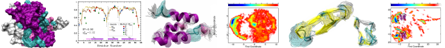
A Highly Coupled Network of Tertiary Interactions in the SAM-I Riboswitch and Their Role in Regulatory Tuning
Publication date: 6 November 2015
Source:Journal of Molecular Biology, Volume 427, Issue 22
Author(s): Christopher Wostenberg, Pablo Ceres, Jacob T. Polaski, Robert T. Batey
RNA folding in vivo is significantly influenced by transcription, which is not necessarily recapitulated by Mg2+-induced folding of the corresponding full-length RNA in vitro. Riboswitches that regulate gene expression at the transcriptional level are an ideal system for investigating this aspect of RNA folding as ligand-dependent termination is obligatorily co-transcriptional, providing a clear readout of the folding outcome. The folding of representative members of the SAM-I family of riboswitches has been extensively analyzed using approaches focusing almost exclusively upon Mg2+ and/or S-adenosylmethionine (SAM)-induced folding of full-length transcripts of the ligand binding domain. To relate these findings to co-transcriptional regulatory activity, we have investigated a set of structure-guided mutations of conserved tertiary architectural elements of the ligand binding domain using an in vitro single-turnover transcriptional termination assay, complemented with phylogenetic analysis and isothermal titration calorimetry data. This analysis revealed a conserved internal loop adjacent to the SAM binding site that significantly affects ligand binding and regulatory activity. Conversely, most single point mutations throughout key conserved features in peripheral tertiary architecture supporting the SAM binding pocket have relatively little impact on riboswitch activity. Instead, a secondary structural element in the peripheral subdomain appears to be the key determinant in observed differences in regulatory properties across the SAM-I family. These data reveal a highly coupled network of tertiary interactions that promote high-fidelity co-transcriptional folding of the riboswitch but are only indirectly linked to regulatory tuning.
Graphical abstract
Categories: Journal Articles
Acidic Residues in the Hfq Chaperone Increase the Selectivity of sRNA Binding and Annealing
Publication date: 6 November 2015
Source:Journal of Molecular Biology, Volume 427, Issue 22
Author(s): Subrata Panja, Andrew Santiago-Frangos, Daniel J. Schu, Susan Gottesman, Sarah A. Woodson
Hfq facilitates gene regulation by small non-coding RNAs (sRNAs), thereby affecting bacterial attributes such as biofilm formation and virulence. Escherichia coli Hfq recognizes specific U-rich and AAN motifs in sRNAs and target mRNAs, after which an arginine patch on the rim promotes base pairing between their complementary sequences. In the cell, Hfq must discriminate between many similar RNAs. Here, we report that acidic amino acids lining the sRNA binding channel between the inner pore and rim of the Hfq hexamer contribute to the selectivity of Hfq's chaperone activity. RNase footprinting, in vitro binding and stopped-flow fluorescence annealing assays showed that alanine substitution of D9, E18 or E37 strengthened RNA interactions with the rim of Hfq and increased annealing of non-specific or U-tailed RNA oligomers. Although the mutants were less able than wild-type Hfq to anneal sRNAs with wild-type rpoS mRNA, the D9A mutation bypassed recruitment of Hfq to an (AAN)4 motif in rpoS, both in vitro and in vivo. These results suggest that acidic residues normally modulate access of RNAs to the arginine patch. We propose that this selectivity limits indiscriminate target selection by E. coli Hfq and enforces binding modes that favor genuine sRNA and mRNA pairs.
Graphical abstract
Categories: Journal Articles
Mechanistic Analysis of Activation of the Innate Immune Sensor PKR by Bacterial RNA
Publication date: 6 November 2015
Source:Journal of Molecular Biology, Volume 427, Issue 22
Author(s): Chelsea M. Hull, Philip C. Bevilacqua
The protein kinase PKR (protein kinase R) is a sensor in innate immunity. PKR autophosphorylates in the presence of double-stranded RNA enabling it to phosphorylate its substrate, eIF2α (eukaryotic initiation factor 2α), halting cellular translation. Classical activators of PKR are long viral double-stranded RNAs, but recently, PKR has been found to be activated by bacterial RNA. However, the features of bacterial RNA that activate PKR are unknown. We studied the Bacillus subtilis trp 5′-UTR (untranslated region), which is an indirect riboswitch with secondary and tertiary RNA structures that regulate gene function. Additionally, the trp 5′-UTR binds a protein, TRAP (tryptophan RNA-binding attenuation protein), which recognizes l-tryptophan. We present the first evidence that multiple structural features in this RNA, which are typical of bacterial RNAs, activate PKR in TRAP-free and TRAP/l-Trp-bound forms. Segments from the 5′-UTR, including the terminator 5′-stem–loop and Shine–Dalgarno blocking hairpins, demonstrated 5′-triphosphate and flanking RNA tail dependence on PKR activation. Disruption of long-distance tertiary interactions in the 5′-UTR led to partial loss in activation, consistent with highly base-paired regions in bacterial RNA activating PKR. One physiological change a bacterial RNA would face in a human cell is a decrease in the concentration of free magnesium. Upon lowering the magnesium concentration to human physiological conditions of 0.5mM, the trp 5′-UTR continued to activate PKR potently. Moreover, total RNA from Escherichia coli, depleted of rRNA, also activated PKR under these ionic conditions. This study demonstrates that PKR can signal the presence of bacterial RNAs under physiological ionic conditions and offers a potential explanation for the apparent absence of riboswitches in the human genome.
Graphical abstract
Categories: Journal Articles
Mutations in RNA Polymerase Bridge Helix and Switch Regions Affect Active-Site Networks and Transcript-Assisted Hydrolysis
Publication date: 6 November 2015
Source:Journal of Molecular Biology, Volume 427, Issue 22
Author(s): Nan Zhang, Jorrit Schäfer, Amit Sharma, Lucy Rayner, Xiaodong Zhang, Roman Tuma, Peter Stockley, Martin Buck
In bacterial RNA polymerase (RNAP), the bridge helix and switch regions form an intricate network with the catalytic active centre and the main channel. These interactions are important for catalysis, hydrolysis and clamp domain movement. By targeting conserved residues in Escherichia coli RNAP, we are able to show that functions of these regions are differentially required during σ70-dependent and the contrasting σ54-dependent transcription activations and thus potentially underlie the key mechanistic differences between the two transcription paradigms. We further demonstrate that the transcription factor DksA directly regulates σ54-dependent activation both positively and negatively. This finding is consistent with the observed impacts of DksA on σ70-dependent promoters. DksA does not seem to significantly affect RNAP binding to a pre-melted promoter DNA but affects extensively activity at the stage of initial RNA synthesis on σ54-regulated promoters. Strikingly, removal of the σ54 Region I is sufficient to invert the action of DksA (from stimulation to inhibition or vice versa) at two test promoters. The RNAP mutants we generated also show a strong propensity to backtrack. These mutants increase the rate of transcript-hydrolysis cleavage to a level comparable to that seen in the Thermus aquaticus RNAP even in the absence of a non-complementary nucleotide. These novel phenotypes imply an important function of the bridge helix and switch regions as an anti-backtracking ratchet and an RNA hydrolysis regulator.
Graphical abstract
Categories: Journal Articles
Three-Dimensional Structure of Vertebrate Muscle Z-Band: The Small-Square Lattice Z-Band in Rat Cardiac Muscle
Publication date: 6 November 2015
Source:Journal of Molecular Biology, Volume 427, Issue 22
Author(s): Thomas Burgoyne, Edward P. Morris, Pradeep K. Luther
The Z-band in vertebrate striated muscle crosslinks actin filaments of opposite polarity from adjoining sarcomeres and transmits tension along myofibrils during muscular contraction. It is also the location of a number of proteins involved in signalling and myofibrillogenesis; mutations in these proteins lead to myopathies. Understanding the high-resolution structure of the Z-band will help us understand its role in muscle contraction and the role of these proteins in the function of muscle. The appearance of the Z-band in transverse-section electron micrographs typically resembles a small-square lattice or a basketweave appearance. In longitudinal sections, the Z-band width varies more with muscle type than species: slow skeletal and cardiac muscles have wider Z-bands than fast skeletal muscles. As the Z-band is periodic, Fourier methods have previously been used for three-dimensional structural analysis. To cope with variations in the periodic structure of the Z-band, we have used subtomogram averaging of tomograms of rat cardiac muscle in which subtomograms are extracted and compared and similar ones are averaged. We show that the Z-band comprises four to six layers of links, presumably α-actinin, linking antiparallel overlapping ends of the actin filaments from the adjoining sarcomeres. The reconstruction shows that the terminal 5–7nm of the actin filaments within the Z-band is devoid of any α-actinin links and is likely to be the location of capping protein CapZ.
Graphical abstract
Categories: Journal Articles
Substrate-Induced Allosteric Change in the Quaternary Structure of the Spermidine N-Acetyltransferase SpeG
Publication date: 6 November 2015
Source:Journal of Molecular Biology, Volume 427, Issue 22
Author(s): Ekaterina V. Filippova, Steven Weigand, Jerzy Osipiuk, Olga Kiryukhina, Andrzej Joachimiak, Wayne F. Anderson
The spermidine N-acetyltransferase SpeG is a dodecameric enzyme that catalyzes the transfer of an acetyl group from acetyl coenzyme A to polyamines such as spermidine and spermine. SpeG has an allosteric polyamine-binding site and acetylating polyamines regulate their intracellular concentrations. The structures of SpeG from Vibrio cholerae in complexes with polyamines and cofactor have been characterized earlier. Here, we present the dodecameric structure of SpeG from V. cholerae in a ligand-free form in three different conformational states: open, intermediate and closed. All structures were crystallized in C2 space group symmetry and contain six monomers in the asymmetric unit cell. Two hexamers related by crystallographic 2-fold symmetry form the SpeG dodecamer. The open and intermediate states have a unique open dodecameric ring. This SpeG dodecamer is asymmetric except for the one 2-fold axis and is unlike any known dodecameric structure. Using a fluorescence thermal shift assay, size-exclusion chromatography with multi-angle light scattering, small-angle X-ray scattering analysis, negative-stain electron microscopy and structural analysis, we demonstrate that this unique open dodecameric state exists in solution. Our combined results indicate that polyamines trigger conformational changes and induce the symmetric closed dodecameric state of the protein when they bind to their allosteric sites.
Graphical abstract
Categories: Journal Articles
Sequence–Structure–Function Classification of a Catalytically Diverse Oxidoreductase Superfamily in Mycobacteria
Publication date: 6 November 2015
Source:Journal of Molecular Biology, Volume 427, Issue 22
Author(s): F. Hafna Ahmed, Paul D. Carr, Brendon M. Lee, Livnat Afriat-Jurnou, A. Elaaf Mohamed, Nan-Sook Hong, Jack Flanagan, Matthew C. Taylor, Chris Greening, Colin J. Jackson
The deazaflavin cofactor F420 enhances the persistence of mycobacteria during hypoxia, oxidative stress, and antibiotic treatment. However, the identities and functions of the mycobacterial enzymes that utilize F420 under these conditions have yet to be resolved. In this work, we used sequence similarity networks to analyze the distribution of the largest F420-dependent protein family in mycobacteria. We show that these enzymes are part of a larger split β-barrel enzyme superfamily (flavin/deazaflavin oxidoreductases, FDORs) that include previously characterized pyridoxamine/pyridoxine-5′-phosphate oxidases and heme oxygenases. We show that these proteins variously utilize F420, flavin mononucleotide, flavin adenine dinucleotide, and heme cofactors. Functional annotation using phylogenetic, structural, and spectroscopic methods revealed their involvement in heme degradation, biliverdin reduction, fatty acid modification, and quinone reduction. Four novel crystal structures show that plasticity in substrate binding pockets and modifications to cofactor binding motifs enabled FDORs to carry out a variety of functions. This systematic classification and analysis provides a framework for further functional analysis of the roles of FDORs in mycobacterial pathogenesis and persistence.
Graphical abstract
Categories: Journal Articles
The Antibody Light-Chain Linker Is Important for Domain Stability and Amyloid Formation
Publication date: 6 November 2015
Source:Journal of Molecular Biology, Volume 427, Issue 22
Author(s): Cardine N. Nokwe, Manuel Hora, Martin Zacharias, Hisashi Yagi, Christine John, Bernd Reif, Yuji Goto, Johannes Buchner
The association of light chains (LCs) and heavy chains is the basis for functional antibodies that are essential for adaptive immune responses. However, in some cases, LCs and especially fragments consisting of the LC variable (VL) domain are pathologically deposited in fatal aggregation diseases. The two domains of the LC are connected by a highly conserved linker. We show here that, unexpectedly, the linker residue Arg108 affects the conformational stability and folding of both VLκ and LC constant (CLκ) domains. Interestingly, the extension of VL by Arg108 results in its resistance to amyloid formation, which suggests that the nature of the truncation of the LC plays a crucial role in disease progression. Increased solvation due to the exposed charged C-terminal Arg108 residue explains its stabilizing effects on the VL domain. For the CL domain, the interaction of N-terminal loop residues with Arg108 is important for the integrity of the domain, as the disruption of this interaction results in fluctuation, partial opening of the protein's interior and the exposure of hydrophobic residues that destabilize the domain. This establishes new principles for antibody domain architecture and amyloidogenicity.
Graphical abstract
Categories: Journal Articles
Effects of Linker Length and Transient Secondary Structure Elements in the Intrinsically Disordered Notch RAM Region on Notch Signaling
Publication date: 6 November 2015
Source:Journal of Molecular Biology, Volume 427, Issue 22
Author(s): Kathryn P. Sherry, Scott E. Johnson, Christine L. Hatem, Ananya Majumdar, Doug Barrick
Formation of the bivalent interaction between the Notch intracellular domain (NICD) and the transcription factor CBF-1/RBP-j, Su(H), Lag-1 (CSL) is a key event in Notch signaling because it switches Notch-responsive genes from a repressed state to an activated state. Interaction of the intrinsically disordered RBP-j-associated molecule (RAM) region of NICD with CSL is thought to both disrupt binding of corepressor proteins to CSL and anchor NICD to CSL, promoting interaction of the ankyrin domain of NICD with CSL through an effective concentration mechanism. To quantify the role of disorder in the RAM linker region on the effective concentration enhancement of Notch transcriptional activation, we measured the effects of linker length variation on activation. The resulting activation profile has general features of a worm-like chain model for effective concentration. However, deviations from the model for short sequence deletions suggest that RAM contains sequence-specific structural elements that may be important for activation. Structural characterization of the RAM linker with sedimentation velocity analytical ultracentrifugation and NMR spectroscopy reveals that the linker is compact and contains three transient helices and two extended and dynamic regions. To test if these secondary structure elements are important for activation, we made sequence substitutions to change the secondary structure propensities of these elements and measured transcriptional activation of the resulting variants. Substitutions to two of these nonrandom elements (helix 2, extended region 1) have effects on activation, but these effects do not depend on the nature of the substituting residues. Thus, the primary sequences of these elements, but not their secondary structures, are influencing signaling.
Graphical abstract
Categories: Journal Articles
Peptide Probes Reveal a Hydrophobic Steric Ratchet in the Anthrax Toxin Protective Antigen Translocase
Publication date: 6 November 2015
Source:Journal of Molecular Biology, Volume 427, Issue 22
Author(s): Jennifer M. Colby, Bryan A. Krantz
Anthrax toxin is a tripartite virulence factor produced by Bacillus anthracis during infection. Under acidic endosomal pH conditions, the toxin's protective antigen (PA) component forms a transmembrane channel in host cells. The PA channel then translocates its two enzyme components, lethal factor and edema factor, into the host cytosol under the proton motive force. Protein translocation under a proton motive force is catalyzed by a series of nonspecific polypeptide binding sites, called clamps. A 10-residue guest/host peptide model system, KKKKKXXSXX, was used to functionally probe polypeptide-clamp interactions within wild-type PA channels. The guest residues were Thr, Ala, Leu, Phe, Tyr, and Trp. In steady-state translocation experiments, the channel blocked most tightly with peptides that had increasing amounts of nonpolar surface area. Cooperative peptide binding was observed in the Trp-containing peptide sequence but not the other tested sequences. Trp substitutions into a flexible, uncharged linker between the lethal factor amino-terminal domain and diphtheria toxin A chain expedited translocation. Therefore, peptide-clamp sites in translocase channels can sense large steric features (like tryptophan) in peptides, and while these steric interactions may make a peptide translocate poorly, in the context of folded domains, they can make the protein translocate more rapidly presumably via a hydrophobic steric ratchet mechanism.
Graphical abstract
Categories: Journal Articles
Functional Monomerization of a ClC-Type Fluoride Transporter
Publication date: 6 November 2015
Source:Journal of Molecular Biology, Volume 427, Issue 22
Author(s): Nicholas B. Last, Christopher Miller
Anion channels and antiporters of the ClC superfamily have been found to be exclusively dimeric in nature, even though each individual monomer contains the complete transport pathway. Here, we describe the destabilization through mutagenesis of the dimer interface of a bacterial F−/H+ antiporter, ClCF-eca. Several mutations that produce monomer/dimer equilibrium of the normally dimeric transporter were found, simply by shortening a hydrophobic side chain in some cases. One mutation, L376W, leads to a wholly monomeric variant that shows full activity. Furthermore, we discovered a naturally destabilized homologue, ClCF-rla, which undergoes partial monomerization in detergent without additional mutations. These results, in combination with the previous functional monomerization of the distant relative ClC-ec1, demonstrate that the monomer alone is the functional unit for several clades of the ClC superfamily.
Graphical abstract
Categories: Journal Articles
Protein–Protein Interfaces in Viral Capsids Are Structurally Unique
Publication date: 6 November 2015
Source:Journal of Molecular Biology, Volume 427, Issue 22
Author(s): Shanshan Cheng, Charles L. Brooks
Viral capsids exhibit elaborate and symmetrical architectures of defined sizes and remarkable mechanical properties not seen with cellular macromolecular complexes. Given the uniqueness of the higher-order organization of viral capsid proteins in the virosphere, we explored the question of whether the patterns of protein–protein interactions within viral capsids are distinct from those in generic protein complexes. Our comparative analysis involving a non-redundant set of 551 inter-subunit interfaces in viral capsids from VIPERdb and 20,014 protein–protein interfaces in non-capsid protein complexes from the Protein Data Bank found 418 generic protein–protein interfaces that share similar physicochemical patterns with some protein–protein interfaces in the capsid set, using the program PCalign we developed for comparing protein–protein interfaces. This overlap in the structural space of protein–protein interfaces is significantly small, with a p-value <0.0001, based on a permutation test on the total set of protein–protein interfaces. Furthermore, the generic protein–protein interfaces that bear similarity in their spatial and chemical arrangement with capsid ones are mostly small in size with fewer than 20 interfacial residues, which results from the relatively limited choices of natural design for small interfaces rather than having significant biological implications in terms of functional relationships. We conclude based on this study that protein–protein interfaces in viral capsids are non-representative of patterns in the smaller, more compact cellular protein complexes. Our finding highlights the design principle of building large biological containers from repeated, self-assembling units and provides insights into specific targets for antiviral drug design for improved efficacy.
Graphical abstract
Categories: Journal Articles
Bacterial Networks in Cells and Communities
Publication date: Available online 24 October 2015
Source:Journal of Molecular Biology
Author(s): Victor Sourjik, Julia A. Vorholt
Research on the bacterial regulatory networks is currently experiencing a true revival, driven by advances in methodology and by emergence of novel concepts. The biannual conference Bacterial Networks (BacNet15) held in May 2015, in Sant Feliu de Guíxols, Spain, covered progress in the studies of regulatory networks that control bacterial physiology, cell biology, stress responses, metabolism, collective behavior and evolution. It demonstrated how interdisciplinary approaches that combine molecular biology and biochemistry with the latest microscopy developments, whole cell (−omics) approaches and mathematical modeling can help understand design principles relevant in microbiology. It further showed how current biotechnology and medical microbiology could profit from our knowledge of and ability to engineer regulatory networks of bacteria.
Graphical abstract
Categories: Journal Articles
Editorial Board
Publication date: 23 October 2015
Source:Journal of Molecular Biology, Volume 427, Issue 21
Source:Journal of Molecular Biology, Volume 427, Issue 21
Categories: Journal Articles
Contents List
Publication date: 23 October 2015
Source:Journal of Molecular Biology, Volume 427, Issue 21
Source:Journal of Molecular Biology, Volume 427, Issue 21
Categories: Journal Articles