
Journal of Molecular Biology
Characterization of the Catalytic Domain of Human APOBEC3B and the Critical Structural Role for a Conserved Methionine
Publication date: Available online 14 August 2015
Source:Journal of Molecular Biology
Author(s): Sachini U. Siriwardena, Thisari A. Guruge, Ashok S. Bhagwat
Human APOBEC3B deaminates cytosines in DNA and belongs to the AID/APOBEC family of enzymes. These proteins are involved in innate and adaptive immunity and may cause mutations in a variety of cancers. To characterize its ability to convert cytosines into uracils, we tested several derivatives of APOBEC3B gene for their ability to cause mutations in Escherichia coli. Through this analysis, a methionine residue at the junction of the amino-terminal domain (NTD) and the carboxy-terminal domain (CTD) was found to be essential for high mutagenicity. Properties of mutants with substitutions at this position, examination of existing molecular structures of APOBEC3 family members and molecular modeling suggest that this residue is essential for the structural stability of this family of proteins. The APOBEC3B CTD with the highest mutational activity was purified to homogeneity and its kinetic parameters were determined. Size-exclusion chromatography of the CTD monomer showed that it is in equilibrium with its dimeric form and matrix-assisted laser desorption ionization time-of-flight analysis of the protein suggested that the dimer may be quite stable. The partially purified NTD did not show intrinsic deamination activity and did not enhance the activity of the CTD in biochemical assays. Finally, APOBEC3B was at least 10-fold less efficient at mutating 5-methylcytosine (5mC) to thymine than APOBEC3A in a genetic assay and was at least 10-fold less efficient at deaminating 5mC compared to C in biochemical assays. These results shed light on the structural organization of APOBEC3B catalytic domain, its substrate specificity and its possible role in causing genome-wide mutations.
Graphical abstract
Categories: Journal Articles
Selection of High-Affinity Peptidic Serine Protease Inhibitors with Increased Binding Entropy from a Back-Flip Library of Peptide–Protease Fusions
Publication date: Available online 14 August 2015
Source:Journal of Molecular Biology
Author(s): Hans Peter Sørensen, Peng Xu, Longguang Jiang, Tobias Kromann-Hansen, Knud J. Jensen, Mingdong Huang, Peter A. Andreasen
We have developed a new concept for designing peptidic protein modulators, by recombinantly fusing the peptidic modulator, with randomized residues, directly to the target protein via a linker and screening for internal modulation of the activity of the protein. We tested the feasibility of the concept by fusing a 10-residue-long, disulfide-bond-constrained inhibitory peptide, randomized in selected positions, to the catalytic domain of the serine protease murine urokinase-type plasminogen activator. High-affinity inhibitory peptide variants were identified as those that conferred to the fusion protease the lowest activity for substrate hydrolysis. The usefulness of the strategy was demonstrated by the selection of peptidic inhibitors of murine urokinase-type plasminogen activator with a low nanomolar affinity. The high affinity could not have been predicted by rational considerations, as the high affinity was associated with a loss of polar interactions and an increased binding entropy.
Graphical abstract
Categories: Journal Articles
β-Structure within the Denatured State of the Helical Protein Domain BBL
Publication date: Available online 14 August 2015
Source:Journal of Molecular Biology
Author(s): Lipi Thukral, Simone Schwarze, Isabella Daidone, Hannes Neuweiler
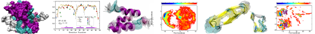
Protein denatured states are the origin of both healthy and toxic conformational species. Denatured states of ultrafast folding proteins are of interest in mechanistic studies because they are energetically close to the kinetic bottleneck of folding. However, their transient nature makes them elusive to experiment. Here, we generated the denatured state of the helical domain BBL that is poised to fold in microseconds by a single-point mutation and combined circular dichroism spectroscopy, single-molecule fluorescence fluctuation analysis, and computer simulation to characterize its structure and dynamics. Circular dichroism showed a largely unfolded ensemble with marginal helix but significant β-sheet content. Main-chain structure and dynamics were unaffected by side-chain interactions that stabilize the native state, as revealed by site-directed mutagenesis and nanosecond loop closure kinetics probed by fluorescence correlation spectroscopy. Replica-exchange and constant-temperature molecular dynamics simulations showed a highly collapsed, hydrogen-bonded denatured state containing turn and β-sheet structure and few nucleating helices in an otherwise unfolded ensemble. An irregular β-hairpin element that connects helices in the native fold was poised to be formed. The surprising observation of β-structure in regions that form helices in the native state is reconciled by a generic low-energy pathway from the northwest quadrant of Ramachandran space to the helical basin present under folding conditions, proposed recently. Our results show that, indeed, rapid nucleation of helix emanates from β-structure formed early within a collapsed ensemble of unfolded conformers.
Graphical abstract
Source:Journal of Molecular Biology
Author(s): Lipi Thukral, Simone Schwarze, Isabella Daidone, Hannes Neuweiler
Protein denatured states are the origin of both healthy and toxic conformational species. Denatured states of ultrafast folding proteins are of interest in mechanistic studies because they are energetically close to the kinetic bottleneck of folding. However, their transient nature makes them elusive to experiment. Here, we generated the denatured state of the helical domain BBL that is poised to fold in microseconds by a single-point mutation and combined circular dichroism spectroscopy, single-molecule fluorescence fluctuation analysis, and computer simulation to characterize its structure and dynamics. Circular dichroism showed a largely unfolded ensemble with marginal helix but significant β-sheet content. Main-chain structure and dynamics were unaffected by side-chain interactions that stabilize the native state, as revealed by site-directed mutagenesis and nanosecond loop closure kinetics probed by fluorescence correlation spectroscopy. Replica-exchange and constant-temperature molecular dynamics simulations showed a highly collapsed, hydrogen-bonded denatured state containing turn and β-sheet structure and few nucleating helices in an otherwise unfolded ensemble. An irregular β-hairpin element that connects helices in the native fold was poised to be formed. The surprising observation of β-structure in regions that form helices in the native state is reconciled by a generic low-energy pathway from the northwest quadrant of Ramachandran space to the helical basin present under folding conditions, proposed recently. Our results show that, indeed, rapid nucleation of helix emanates from β-structure formed early within a collapsed ensemble of unfolded conformers.
Graphical abstract
Categories: Journal Articles
Shelter in a swarm
Publication date: Available online 12 August 2015
Source:Journal of Molecular Biology
Author(s): Rasika M. Harshey, Jonathan D. Partridge
Flagella propel bacteria during both swimming and swarming, dispersing them widely. However, while swimming bacteria use chemotaxis to find nutrients and avoid toxic environments, swarming bacteria appear to suppress chemotaxis and to use the dynamics of their collective motion to continuously expand and acquire new territory, barrel through lethal chemicals in their path, carry along bacterial and fungal cargo that assists in exploration of new niches, and engage in group warfare for niche dominance. Here we focus on two aspects of swarming, which if understood, hold the promise of revealing new insights into microbial signaling and behavior, with ramifications beyond bacterial swarming. These are: how bacteria sense they are on a surface and turn on programs that promote movement, and how as dense packs they override scarcity and adversity.
Graphical abstract
Categories: Journal Articles
Analysis of a FANCE Splice Isoform in Regard to DNA Repair
Publication date: Available online 12 August 2015
Source:Journal of Molecular Biology
Author(s): Frédérick Bouffard, Karine Plourde, Simon Bélanger, Geneviève Ouellette, Yvan Labrie, Francine Durocher
The FANC-BRCA DNA repair pathway is activated in response to interstrand crosslinks formed in DNA. A homozygous mutation in 1 of the 17 Fanconi anemia (FA) genes results in malfunctions of this pathway and development of FA syndrome. The integrity of this protein network is essential for good maintenance of DNA repair process and genome stability. Following the identification of an alternatively splice isoform of FANCE (Fanconi anemia complementation group E) significantly expressed in breast cancer individuals from high-risk non-BRCA1/2 families, we studied the impact of this FANCE splice isoform (FANCEΔ4) on DNA repair processes. We have demonstrated that FANCEΔ4 mRNA was efficiently translated into a functional protein and expressed in normal and breast cancer cell lines. Following treatment with the crosslinking agent mitomycin C, EUFA130 (FANCE-deficient) cells infected with FANCEΔ4 were blocked into G2/M phase, while cell survival was significantly reduced compared with FANCE-infected EUFA130 cells. In addition, FANCEΔ4 did not allow FANCD2 and FANCI monoubiquitination, which represents a crucial step of the FANC-BRCA functional pathway. As observed for FANCE wild-type protein, localization of FANCEΔ4 protein was confined to the nucleus following mitomycin C treatment. Although FANCEΔ4 protein showed interaction with FANCE, FANCEΔ4 did not support normal function of FANCE protein in this pathway and could have deleterious effects on FANCE protein activity. We have demonstrated that FANCEΔ4 seems to act as a regulator of FANCD2 protein expression level by promoting its degradation. This study highlights the importance of an efficient regulation of alternative splicing expression of FA genes for proper DNA repair.
Graphical abstract
Categories: Journal Articles
DULIP: A dual luminescence-based co-immunoprecipitation assay for interactome mapping in mammalian cells
Publication date: Available online 8 August 2015
Source:Journal of Molecular Biology
Author(s): Philipp Trepte, Alexander Buntru, Konrad Klockmeier, Lindsay Willmore, Anup Arumughan, Christopher Secker, Martina Zenkner, Lydia Brusendorf, Kirstin Rau, Alexandra Redel, Erich E. Wanker
Mapping of protein-protein interactions (PPIs) is critical for understanding protein function and complex biological processes. Here, we present DULIP, a dual luminescence-based co-immunoprecipitation assay, for systematic PPI mapping in mammalian cells. DULIP is a second-generation luminescence-based PPI screening method for the systematic and quantitative analysis of co-immunoprecipitations using two different luciferase tags. Benchmarking studies with positive and negative PPI reference sets revealed that DULIP allows the detection of interactions with high sensitivity and specificity. Furthermore, the analysis of a PPI reference set with known binding affinities demonstrated that both low- and high-affinity interactions can be detected with DULIP assays. Finally, using the well-characterized interaction between Syntaxin-1 and Munc18, we found that DULIP is capable of detecting the effects of point mutations on interaction strength. Taken together, our studies demonstrate that DULIP is a sensitive and reliable method of great utility for systematic interactome research. It can be applied for interaction screening as well as for the validation of PPIs in mammalian cells. Moreover, DULIP permits the specific analysis of mutation-dependent binding patterns.
Graphical abstract
Categories: Journal Articles
The Telomere Binding Protein Cdc13 and the Single-Stranded DNA Binding Protein RPA Protect Telomeric DNA from Resection by Exonucleases
Publication date: Available online 8 August 2015
Source:Journal of Molecular Biology
Author(s): Matthew Greetham, Emmanuel Skordalakes, David Lydall, Bernard A. Connolly
The telomere is present at the ends of all eukaryotic chromosomes and usually consists of repetitive TG-rich DNA that terminates in a single-stranded 3′ TG extension and a 5′ CA-rich recessed strand. A biochemical assay that allows the in vitro observation of exonuclease-catalyzed degradation (resection) of telomeres has been developed. The approach uses an oligodeoxynucleotide that folds to a stem–loop with a TG-rich double-stranded region and a 3′ single-stranded extension, typical of telomeres. Cdc13, the major component of the telomere-specific CST complex, strongly protects the recessed strand from the 5′→3′ exonuclease activity of the model exonuclease from bacteriophage λ. The isolated DNA binding domain of Cdc13 is less effective at shielding telomeres. Protection is specific, not being observed in control DNA lacking the specific TG-rich telomere sequence. RPA, the eukaryotic single-stranded DNA binding protein, also inhibits telomere resection. However, this protein is non-specific, equally hindering the degradation of non-telomere controls.
Graphical abstract
Categories: Journal Articles
The LcrG Tip Chaperone Protein of the Yersinia pestis Type III Secretion System Is Partially Folded
Publication date: Available online 7 August 2015
Source:Journal of Molecular Biology
Author(s): Sukanya Chaudhury, Clarice de Azevedo Souza, Gregory V. Plano, Roberto N. De Guzman
The type III secretion system (T3SS) is essential in the pathogenesis of Yersinia pestis, the causative agent of plague. A small protein, LcrG, functions as a chaperone to the tip protein LcrV, and the LcrG–LcrV interaction is important in regulating protein secretion through the T3SS. The atomic structure of the LcrG family is currently unknown. However, because of its predicted helical propensity, many have suggested that the LcrG family forms a coiled-coil structure. Here, we show by NMR and CD spectroscopy that LcrG lacks a tertiary structure and it consists of three partially folded α-helices spanning residues 7–38, 41–46, and 58–73. NMR titrations of LcrG with LcrV show that the entire length of a truncated LcrG (residues 7–73) is involved in binding to LcrV. However, there is regional variation in how LcrG binds to LcrV. The C-terminal region of a truncated LcrG (residues 52–73) shows tight binding interaction with LcrV while the N-terminal region (residues 7–51) shows weaker interaction with LcrV. This suggests that there are at least two binding events when LcrG binds to LcrV. Biological assays and mutagenesis indicate that the C-terminal region of LcrG (residues 52–73) is important in blocking protein secretion through the T3SS. Our results reveal structural and mechanistic insights into the atomic conformation of LcrG and how it binds to LcrV.
Graphical abstract
Categories: Journal Articles
Gene Regulation Gets in Tune: How Riboswitch Tertiary-Structure Networks Adapt to Meet the Needs of Their Transcription Units
Publication date: Available online 6 August 2015
Source:Journal of Molecular Biology
Author(s): Debapratim Dutta, Joseph E. Wedekind
Source:Journal of Molecular Biology
Author(s): Debapratim Dutta, Joseph E. Wedekind
Categories: Journal Articles
Influence of Internal DNA Pressure on Stability and Infectivity of Phage λ
Publication date: Available online 5 August 2015
Source:Journal of Molecular Biology
Author(s): D.W. Bauer, A. Evilevitch
Viruses must remain infectious while in harsh extracellular environments. An important aspect of viral particle stability for double-stranded DNA viruses is the energetically unfavorable state of the tightly confined DNA chain within the virus capsid creating pressures of tens of atmospheres. Here, we study the influence of internal genome pressure on the thermal stability of viral particles. Using differential scanning calorimetry to monitor genome loss upon heating, we find that internal pressure destabilizes the virion, resulting in a smaller activation energy barrier to trigger DNA release. These experiments are complemented by plaque assay and electron microscopy measurements to determine the influence of intra-capsid DNA pressure on the rates of viral infectivity loss. At higher temperatures (65–75°C), failure to retain the packaged genome is the dominant mechanism of viral inactivation. Conversely, at lower temperatures (40–55°C), a separate inactivation mechanism dominates, which results in non-infectious particles that still retain their packaged DNA. Most significantly, both mechanisms of infectivity loss are directly influenced by internal DNA pressure, with higher pressure resulting in a more rapid rate of inactivation at all temperatures.
Graphical abstract
Categories: Journal Articles